Pathology
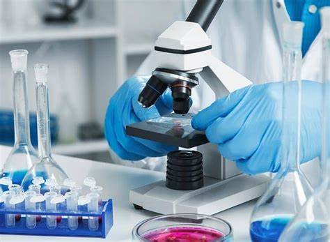
Pathology is the medical department in which the patients are examined using their blood, urine, tissues, or other cerebrospinal fluids. Pathologists determine the cause and nature of the illness. We have the best pathologists and they examine the patient properly and give accurate results and guidelines to the patients. In Bhagwandas hospital, you’ll find the best pathologists in Sonipat who will analyze you very firmly and finely.

Our Speciality
Here are some key aspects of pathology:
Diagnostic Pathology: Pathologists analyze tissue samples (biopsies) and examine cells under a microscope, use professional techniques to detect abnormalities, and provide detailed reports to help guide treatment decisions.
Surgical Pathology: Pathologists examine tissue samples removed during surgeries to determine the presence and extent of disease and provide information that helps guide further treatment options.
Cytopathology: Cytopathologists study cells obtained through techniques such as fine needles or Pap smears. They examine these cells microscopically to detect cancer, infections, and other diseases.
In Bhagwan das hospital, you’ll find the best pathologists in Sonipat who will analyze you very firmly and finely.
PROCESS OF PATHOLOGY:
1 ) Specimen Collection: The first step in pathology is the collection of specimens from the patient. This can include blood samples, tissue biopsies, surgical resections, body fluids (such as urine or cerebrospinal fluid), or other samples depending on the suspected disease or condition.
2) Specimen Processing: Once collected, the specimens are transported to the pathology laboratory for processing. The processing methods may vary depending on the type of specimen and the tests required. For example, tissue specimens are typically fixed in formalin, embedded in paraffin wax, and sliced into thin sections for microscopic examination.
3) Macroscopic Examination: In the case of surgical specimens or larger tissue samples, a macroscopic examination is performed. This involves visual inspection and examination of the specimen for any abnormalities, such as tumors, lesions, or structural changes.
4) Microscopic Examination: Microscopic examination is a critical step in pathology. Thin sections of tissues are mounted on glass slides, stained with various dyes, and examined under a microscope.
Pathologists analyze the cellular and tissue morphology to identify any abnormalities or disease-related changes.
5) Ancillary Tests: In addition to microscopic examination, ancillary tests may be performed to further characterize the specimen. These tests can include immunohistochemistry, molecular testing, cytogenetics, flow cytometry, or other specialized techniques. These tests help in identifying specific markers, genetic alterations, or other molecular features associated with certain diseases.
6) Interpretation and Diagnosis: Based on the findings from macroscopic and microscopic examinations, as well as any additional tests, the pathologist makes a diagnosis. They interpret the results, correlate them with the patient’s clinical history, and provide a detailed report of their findings to the referring
physician or healthcare provider.
PROCESS OF PATHOLOGY:
1 ) Specimen Collection: The first step in pathology is the collection of specimens from the patient. This can include blood samples, tissue biopsies, surgical resections, body fluids (such as urine or cerebrospinal fluid), or other samples depending on the suspected disease or condition.
2) Specimen Processing: Once collected, the specimens are transported to the pathology laboratory for processing. The processing methods may vary depending on the type of specimen and the tests required. For example, tissue specimens are typically fixed in formalin, embedded in paraffin wax, and
sliced into thin sections for microscopic examination.
3) Macroscopic Examination: In the case of surgical specimens or larger tissue samples, a macroscopic examination is performed. This involves visual inspection and examination of the specimen for any abnormalities, such as tumors, lesions, or structural changes.
4) Microscopic Examination: Microscopic examination is a critical step in pathology. Thin sections of tissues are mounted on glass slides, stained with various dyes, and examined under a microscope.
Pathologists analyze the cellular and tissue morphology to identify any abnormalities or disease-related changes.
5) Ancillary Tests: In addition to microscopic examination, ancillary tests may be performed to further characterize the specimen. These tests can include immunohistochemistry, molecular testing, cytogenetics, flow cytometry, or other specialized techniques. These tests help in identifying specific markers, genetic alterations, or other molecular features associated with certain diseases.
6) Interpretation and Diagnosis: Based on the findings from macroscopic and microscopic examinations, as well as any additional tests, the pathologist makes a diagnosis. They interpret the results, correlate them with the patient’s clinical history, and provide a detailed report of their findings to the referring
physician or healthcare provider.
7) Consultation and Communication: Pathologists often collaborate with other healthcare professionals, such as oncologists, surgeons, or radiologists, to discuss findings, provide additional insights, or contribute to the overall management of the patient. Clear and timely communication of pathology results to the referring physician is crucial for appropriate patient care and treatment decisions.
8) Quality Assurance and Quality Control: Pathology laboratories have strict quality assurance and quality control measures in place to ensure accuracy and reliability of results. This includes adherence to standardized protocols, proficiency testing, external quality assessment programs, regular equipment maintenance, and ongoing training and education for laboratory staff.
9) Research and Advancements: Pathology also plays a vital role in research and the advancement of medical knowledge. Pathologists contribute to scientific studies, participate in clinical trials, and continuously update their understanding of disease mechanisms and diagnostic techniques.
WHICH HUMAN FLUIDS USED BY PATHOLOGY?
A) Blood: Blood is one of the most frequently analyzed fluids in pathology. It can be obtained through venipuncture or other methods. Blood tests, such as complete blood count (CBC), blood chemistry analysis, coagulation studies, and blood typing, provide important information about the overall health of
a patient, as well as specific diseases or conditions.
B) Urine: Urine is another commonly tested fluid in pathology. Urinalysis can provide information about kidney function, urinary tract infections, the presence of substances or metabolites, and other conditions. Urine samples are often collected in sterile containers and analyzed for various parameters, including pH, specific gravity, protein, glucose, red and white blood cells, and bacteria.
C) Cerebrospinal Fluid (CSF): CSF is the fluid that surrounds the brain and spinal cord. Analysis of CSF is performed through a procedure called lumbar puncture or spinal tap. CSF analysis helps diagnose conditions affecting the central nervous system, such as meningitis, encephalitis, multiple sclerosis, and certain types of tumors.
D) Synovial Fluid: Synovial fluid is found in joints, such as the knees, shoulders, and wrists. Analysis of synovial fluid can aid in the diagnosis of joint-related conditions, including arthritis, infections, and inflammation. It helps evaluate the presence of inflammatory markers, crystals, and the overall integrity of the joint.
E ) Amniotic Fluid: Amniotic fluid surrounds the fetus during pregnancy. It can be obtained through amniocentesis or other procedures. Analysis of amniotic fluid helps evaluate fetal well-being, genetic disorders, and fetal lung maturity.
