Radiology
Radiology and Services
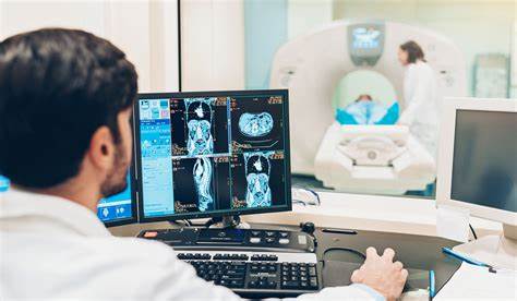
Radiology is a medical facility that is concerned with imaging techniques to examine and treat diseases. Radiologists are medical facilities that are experts in radiology techniques and are familiar with the usage of imaging technologies to interpret diseases.
Medical Imaging Techniques: Radiologists utilize different imaging modalities to visualize and examine various parts of the body. These modalities include:
X-RAY
X-ray imaging is a common diagnostic technique used in medical imaging to produce images of the inside of the body. It is a form of electromagnetic radiation that can pass through tissues and create images based on the differences in the absorption of X-rays by various structures.
Procedure: During an X-ray procedure, the patient is positioned between an X-ray machine and a special film or digital detector. The X-ray machine emits a controlled amount of X-rays that pass through the body and interact differently with various tissues. The X-rays that pass through the body are detected on the other side by the film or digital detector, creating an image.
Types of X-rays: There are various types of X-ray imaging techniques, depending on the area of the body being examined and the information required. Some common types include:
Chest X-ray: This type of X-ray is used to examine the chest area, including the lungs, heart, ribs, and surrounding structures. It can help detect conditions such as pneumonia, lung tumors, collapsed lungs, rib fractures, and heart abnormalities.
Skeletal X-ray: Skeletal X-rays are used to visualize bones and joints throughout the body. They can detect fractures, bone infections, arthritis, bone tumors, and other skeletal abnormalities.
Abdominal X-ray: Abdominal X-rays provide a basic assessment of the organs within the abdomen, including the stomach, liver, intestines, and kidneys. They can help identify conditions such as bowel obstruction, kidney stones, abdominal masses, or the presence of foreign objects.
Dental X-ray: Dental X-rays are specifically used to examine teeth, jawbones, and surrounding structures. They help identify cavities, tooth decay, gum disease, impacted teeth, and other dental conditions.
Spinal X-ray: Spinal X-rays are used to evaluate the bones and alignment of the spine. They can help diagnose spinal fractures, degenerative changes, scoliosis, spinal tumors, or other abnormalities affecting the spine.
Extremity X-ray: Extremity X-rays focus on specific areas of the body, such as the hands, wrists, arms, legs, or feet. They help diagnose fractures, dislocations, joint diseases (e.g., arthritis), or abnormalities in the bones of the extremities.
USG stands for Ultrasonography, also known as ultrasound imaging . It is a technique that uses high- frequency sound waves to produce real-time images of the body’s internal structures.
Abdominal Ultrasound: This type of USG is used to examine the organs in the abdomen, including the liver, gallbladder, pancreas, spleen, kidneys, and intestines. It can help diagnose conditions such as gallstones, liver diseases, kidney stones, abdominal masses, and abnormalities in the gastrointestinal
tract.
Obstetric Ultrasound: Obstetric USG is used during pregnancy to monitor the development and health of the fetus. It can provide information about the gestational age, position of the baby, detect any abnormalities, and assess the placenta and amniotic fluid.
Pelvic Ultrasound: Pelvic USG is performed to examine the reproductive organs in women, including the uterus, ovaries, fallopian tubes, and cervix. It can help diagnose conditions such as ovarian cysts, uterine fibroids, ectopic pregnancies, and abnormalities in the pelvic region.
Transvaginal Ultrasound: This type of USG involves inserting a specially designed transducer into the vagina to obtain detailed images of the pelvic organs, especially the uterus and ovaries. It is commonly used in gynecology for evaluating fertility issues, monitoring early pregnancy, and diagnosing gynecological conditions.
Vascular Ultrasound: Vascular USG is used to assess blood vessels and blood flow. It can help diagnose conditions such as deep vein thrombosis (DVT), peripheral artery disease, aneurysms, or evaluate the blood flow in organs and tissues.
Breast Ultrasound: Breast USG is often used as a complementary imaging technique to mammography for evaluating breast abnormalities. It can help identify cysts, solid masses, or assist in guiding breast biopsies.
Musculoskeletal Ultrasound: Musculoskeletal USG is used to evaluate muscles, tendons, ligaments, and joints. It can aid in diagnosing conditions such as tendon tears, ligament sprains, joint inflammation, and cysts.
Cardiac Ultrasound (Echocardiography): Echocardiography is used to evaluate the structure and function of the heart. It can assess the heart’s chambers, valves, blood flow, and detect any abnormalities or heart conditions.
MRI stands for Magnetic Resonance Imaging. It is a medical imaging technique that uses a strong magnetic field and radio waves to generate detailed images of the internal structures of the body. MRI provides cross-sectional images of organs, tissues, and structures from various angles, allowing for the visualization of both anatomical and functional information.
Here are some common types of MRI techniques:
T1-weighted MRI: T1-weighted MRI provides excellent anatomical detail, highlighting the differences in tissue characteristics. It is often used to visualize the brain, spine, muscles, and other soft tissues. T1-
weighted images show fat as bright and fluid-filled spaces as dark.
T2-weighted MRI: T2-weighted MRI is sensitive to the water content in tissues, making it useful in detecting edema, inflammation, and fluid-filled structures. T2-weighted images show fluid as bright and fat as dark.
FLAIR (Fluid-Attenuated Inversion Recovery): FLAIR MRI is a specialized technique that suppresses the signal from fluid, allowing better visualization of lesions and abnormalities in the brain, particularly those
involving white matter.
Functional MRI (fMRI): fMRI measures and maps changes in blood flow and oxygenation levels in the brain in response to specific tasks or stimuli. It is used to study brain function and can help localize areas involved in speech, motor control, memory, and sensory processing.
Spectroscopy: MRI spectroscopy measures the chemical composition of tissues by analyzing the signals emitted by different molecules. It is used to study brain metabolism, detect brain tumors, and evaluate certain metabolic disorders.